Magnetic Resonance Histology
- Charles E. Putman University, Durham NC, United States
Webster defines "histology" as the microscopic study of tissue structure. Magnetic resonance histology (MRH) was first proposed in 1993 [1]. MR histology has four attributes that distinguish it from traditional optical methods: 1) MRH is nondestructive; 2) MRH probes tissue structure based on "proton stains", i.e. water and how it is bound in the tissue; 3) MRH is 3 dimensional; 4) MRH is inherently digital. This talk will discuss the technical infrastructure required to perform MR histology and demonstrate several applications. The technology has advanced to the point where subtle and fascinating studies of tissue architecture are now possible. We will focus on the use of MRH in neuroscience describing the unique cytoarchitecture that can be seen with conventional gradient echo images, susceptibility images and diffusion tensor imaging.

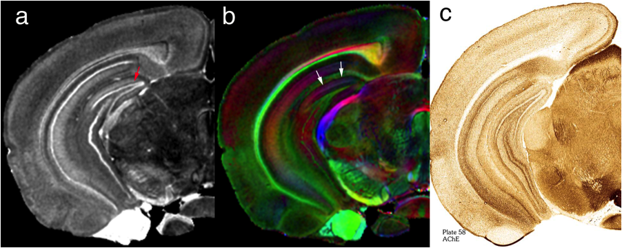
Figure 1 shows selected coronal slices from 3D MR histology images of a mouse brain acquired at 25 um isotropic spatial resolution. Figure 1 a shows the axial diffusivity image and b) shows the color fractional anisotropy image. Figure1 c) shows the acetyl cholinesterase (plate 58) from the classic atlas of the mouse brain based on conventional optical histology [2]. The dentate gyrus, a cell dense region in the hippocampus is clearly seen (red arrow) in figure 1a. Figure 1 b, of the very same tissue at the very same location, reveals completely different cytoarchitecture. Note the subtle shading in the molecular layer demonstrating the radial orientation of the cells (white arrows). The boundaries of subthalamic nuclei are clear in both MR images. Since these are 3D images, volume measures of these sub regions are now routine.
