Diffusion sensitivity of high-resolution 3D TSE in a clinical 3 T system: implications for peripheral nerve imaging
- 1. Technische Universität München, Department of Diagnostic and Interventional Radiology, Munich, Germany
- 2. Philips Healthcare, Hamburg, Germany
- 3. Technische Universität München, Zentralinstitut für Medizintechnik, Garching, Germany
Purpose: 3D turbo-spin-echo (TSE) is a robust technique for fast in vivo high-resolution isotropic T2-weighted imaging. As the 3D TSE voxel size decreases, diffusion weighting (DW) accumulates in the read-out (RO) axis along the flip angle (FA) train. The diffusion sensitivity of 3D TSE has been theoretically addressed based on the extended phase graph (EPG) concept and has been considered practically relevant at clinical systems only when scanning small animals [R1]. However, the 3D TSE voxel size needs to be considerably reduced when fine anatomical structures as peripheral nerves are imaged [R2]. Since nerve diffusion is anisotropic, the diffusion sensitivity of 3D TSE can be dependent on the RO axis orientation. The purpose of the present work is (i) to theoretically investigate the magnitude of diffusion effects in 3D TSE peripheral nerve imaging and (ii) to assess these effects in vivo.
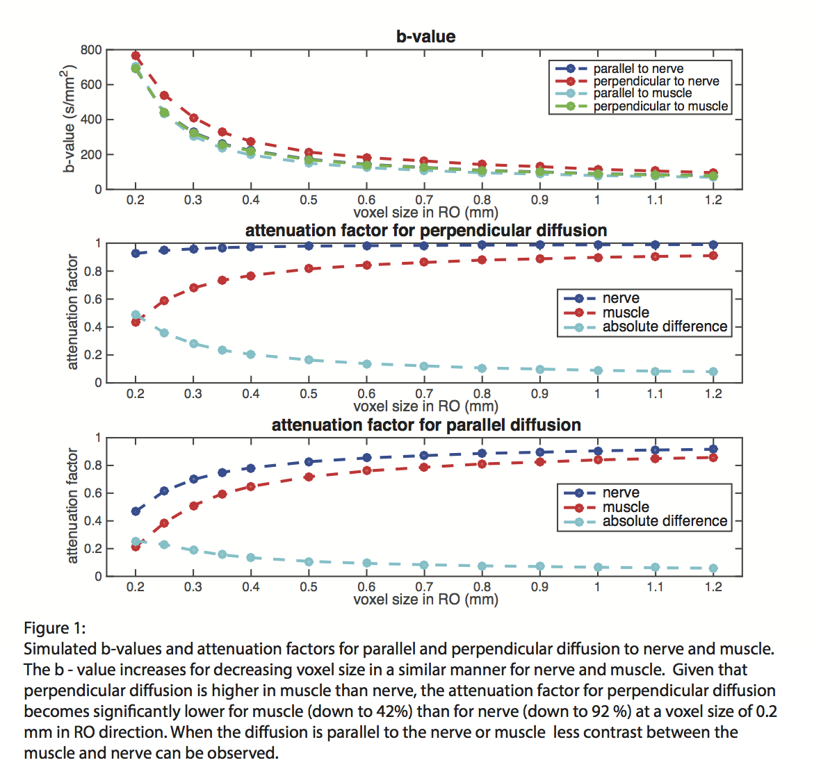
Methods: Simulations were performed for muscle/nerve using RO voxel sizes between 0.2 to 1.2 mm, parallel (PL)/perpendicular (PP) to the long axis of the tissues.
The diffusion operator was introduced in the EPG formulation:
D(k,Δt)=e-D*Δt*k^2, with k(t)=∫ G(t' )dt' and D=Diff. constant.
The attenuation factor A was defined as the ratio of the signal at k = 0 with and without diffusion and the b-value was calculated: b=1/(D*ln(A))
TSE imaging of one healthy subject's knee was performed on a 3T Philips system.
The RO direction was applied PL and PP to the leg axis with FOV = 14×14×8 cm3, TR/TE = 2 s/455 ms, TSE factor = 140, acquisition voxel = 0.5×0.3×3 mm3.
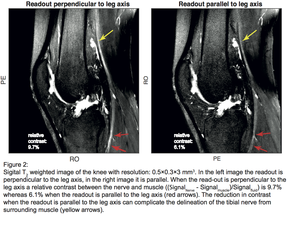
Results: Simulations showed that diffusion effects are stronger for smaller voxels and for RO direction PL to muscle/nerve (Fig. 1). Given that diffusion restriction is stronger in nerve than muscle, the muscle/nerve signal difference decreases when RO direction changes from PP to PL relative to muscle/nerve (Fig. 1). Fig. 2 shows in vivo a 37% reduction in the muscle/tibial nerve contrast when the RO direction changes from PP to PL relative to the leg axis.
Conclusion: Diffusion effects can become relevant in high-resolution 3D TSE imaging of peripheral nerves in a clinical 3T system. Such effects should be considered in FA calculations and protocol optimization.
Acknowledgement: The present work was supported by Philips Healthcare.
- [R1] Weigel Matthias, (2015), Extended phase graphs: Dephasing, RF pulses, and echoes - pure and simple, Journal of Magnetic Resonance Imaging, Volume 41, Issue 2, , 266-295
- [R2] S.K. Thawait et al., (2012), Peripheral Nerve Surgery: The Role of High- Resolution MR Neurography, American Kournal of Neuroradiology Volume:33, 203-210