Nuclear magnetic signal generation with variable field amplitudes
- University of Würzburg, Experimental Physics V, Würzburg, Germany
Introduction
Magnetic Particle Imaging (MPI) is a promising tomographic method, which can determine the distribution of superparamagnetic iron-oxide nanoparticles (SPIOs) directly [1]. Unlike Magnetic Resonance Imaging (MRI), it cannot provide anatomical background information.
To overcome this issue, different approaches have been presented [2]. Due to the different magnetic field configurations required for MPI and MRI, the experiments have to be executed successively.
In this work a novel concept is theoretically investigated to acquire a nuclear induction signal during an MPI measurement.
Methods
For MPI a field free point (FFP) with a strong gradient, generated by a Maxwell coil pair, is moved through the sample. This causes a magnetization change in the SPIOs which can be used for a full 3D reconstruction.

The behavior of the nuclear magnetization vector during a MPI-measurement was numerically analyzed using the Bloch equations and a simplified model of the TWMPI-system. A coil generates an oscillating main magnetic field which cancels out the nuclear magnetization for typical relaxation times and driving frequencies.
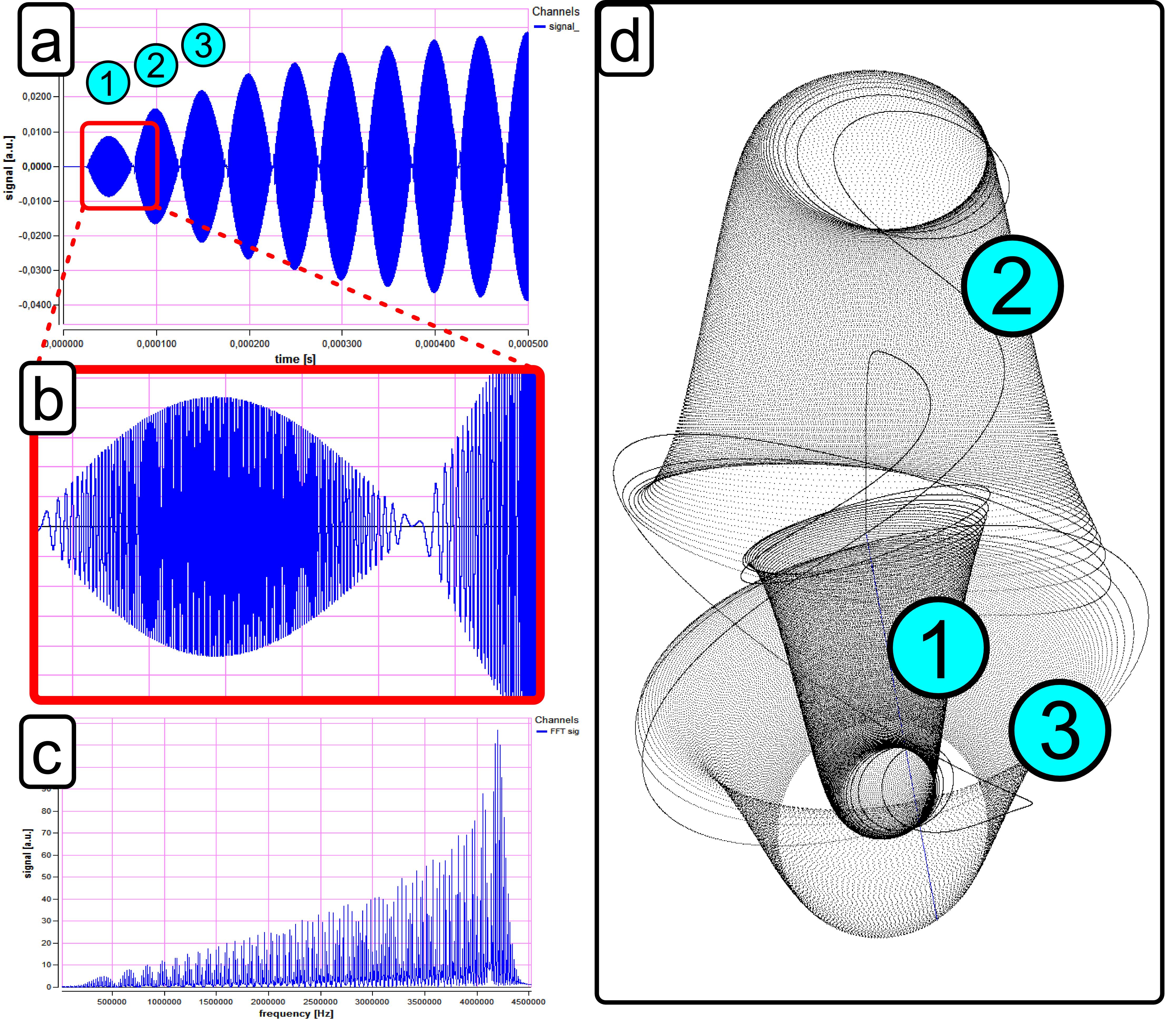
However, applying a perpendicular constant offset field with only 5 percent of the main field strength, a permanent nuclear magnetization arises. In fig. 1 (a) the simulated signal can be seen, which increases according to longitudinal relaxation. A zoom into the signal reveals the frequency sweep (fig. 1 (b)), which can also be seen in the spectrum (fig. 1 (c)). Fig. 1 (d) shows a 3D plot of a magnetization vector over a short time span of two periods.
The zero crossing of the main magnetic field corresponds to the passage of the FFP in the TWMPI-experiment, hence, using the point-by-point scanning method enables spatial encoding.
Conclusion
A novel approach for acquiring a nuclear induction signal has been presented. Contrary to NMR it uses a strong sinusoidal magnetic field and a small constant offset field for magnetization and signal generation. Thus it can be used in a TWMPI-scanner for simultaneously signal acquisition. A closer look at the underlying effect gives an idea about the dynamics of the spin system, which can yield access to measuring new system parameters and may constitute the basis for a new tomographic technique using dynamic magnetic fields.
Funding
DFG: (BE 5293/1-1), EU:project reference 279288
