Integrated TWMPI-MRI scanner - NMR results
- University of Würzburg, Experimental Physics V, Würzburg, Germany
Introduction: Magnetic Particle Imaging (MPI) [1] directly detects superparamagnetic iron-oxide nanoparticles via their nonlinear behavior in alternating fields, while paramagnetic or diamagnetic materials like tissue generate no signal. This leads to a high contrast at the cost of only particles being detectable but not their environment, e.g. the anatomical background. One approach is to combine MPI data with MRI data from a hybrid scanner [2]. More efficiently, MRI and MPI are combined in one scanner using common parts, which is a current research topic [3]. This work presents the basis of a highly integrated TWMPI-MRI scanner.
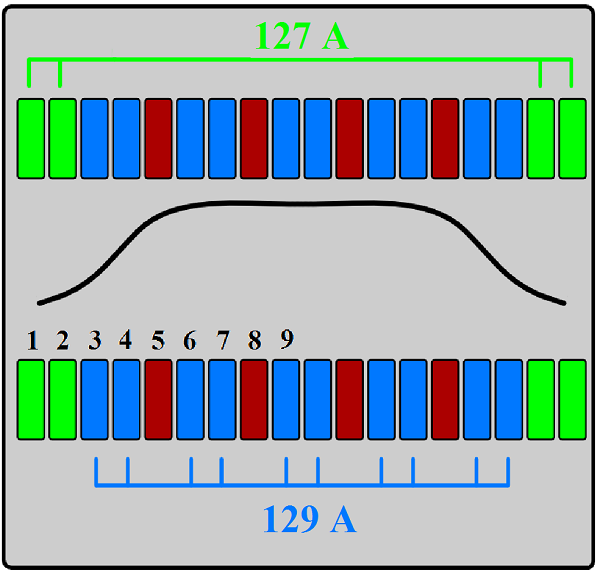
Material and Methods: The most important part of the Traveling Wave MPI scanner [4] is the dynamic Linear Gradient Array (dLGA) which consists of 20 copper coil elements. Each of these coils can be driven individually. The dLGA is used to create a strong magnetic field gradient of about 4 T/m which is necessary for MPI encoding and measurements. For MRI measurements a homogenous magnetic field is required. Using the same coil elements of the dLGA it is also possible to create this field with a tolerance of better than 10-3 in the center [5]. For this purpose strong currents run through 16 out of the 20 coil elements (see fig. 1 green and blue elements). The other four coils (red) are not used for the B0 generation but for the gradient generation in the z-direction. The magnetic field strength was chosen to 235 mT which corresponds to a 1H-Larmor frequency of 10 MHz. A home-built high precision current control unit stabilizes the currents in the dLGA with a precision better than 10-4. As console for the MRI system a further development of a pocket-size NMR device [6] is employed. It can be controlled by a computer running either the device's native pulse program language or Matlab (The Mathworks®). All the hardware is integrated in one circuit board and communications to the microcontroller 68HC908GP32 are possible via USB port. For the pulse shape and the gradients digital-analog converters are used. It operates with a dwell time of 10 µs and therefore a bandwidth of 100 kHz.

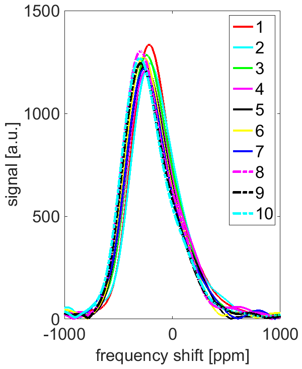
Results: A spin echo experiment has been successfully performed on an oil sample within the B0 field generated by the dLGA. So far only air flow is used for cooling the dLGA, which does not allow continuous operation. The dissipated heat is on the order of 5 kW. For an echo the current was turned on for 350 ms in total to polarize the sample. After that the pulses were applied (echo time of 2 ms) and the echo was acquired. To acquire more than one echo the system was turned on and off with a repetition time of 15 s. The spectra of consecutively acquired echoes show a reproducible Larmor frequency (see fig. 2). The next step will be the encoding along the symmetry axis. Simulations show that coils of the dLGA not used for the B0 field generation can be employed to create a linear gradient over a length of 30 mm with a deviation of less than 1%.
Conclusion: It has been shown that the TWMPI hardware, especially the dLGA, can be used to perform NMR measurements as well. The high precision current control unit is able to maintain a stable field over the whole acquisition time. Even after turning it off and on the magnetic field strength was reproducible. Simulations show that the dLGA can in addition generate a linear gradient over the whole FOV along the system´s main axis.

Acknowledgements: This work was partially funded by the DFG (BE 5293/1-1).
[1] [2] [3] [4] [5] [6]
Introduction: Magnetic Particle Imaging (MPI) [1] directly detects superparamagnetic iron-oxide nanoparticles via their nonlinear behavior in alternating fields while paramagnetic or diamagnetic materials like tissue generate no signal. This leads to a high contrast at the cost of only the particles being detectable but not their environment, e.g. the anatomical background. One approach is to combine MPI data with MRI data from a hybrid scanner [2]. More efficiently, MRI and MPI are combined in one scanner using common parts, which is a current research topic [3]. This work presents the basis of a highly integrated TWMPI-MRI scanner.
Material and Methods: The most important part of the Traveling Wave MPI scanner [4] is the dynamic Linear Gradient Array (dLGA) which consists of 20 copper coil elements. Each of these coils can be driven individually. The dLGA is used to create a strong magnetic field gradient which is necessary for MPI measurements. For MRI measurements a homogenous magnetic field is required. Using the same coil elements it is also possible to create this field with a tolerance of better than 10-3 in the center [5]. The magnetic field strength was chosen to 235 mT corresponding to a 1H-Larmor frequency of 10 MHz. A home-built high precision current control unit stabilizes the currents in the dLGA. As console for the MRI system a further development of a pocket-size NMR device [6] is employed.
Results: A spin echo experiment has been successfully performed on an oil sample within the B0 field generated by the dLGA. The spectra of 10 consecutively acquired echoes show a reproducible Larmor frequency (see fig. 1). The next step will be the encoding along the symmetry axis. Simulations show that coils of the dLGA not used for the B0 field generation can be employed to create a linear gradient over a length of 30 mm.
Conclusion: It has been shown that the TWMPI hardware, especially the dLGA, can be used to perform NMR measurements as well. The high precision current control unit is able to maintain a stable field over the whole acquisition time. Simulations show that the dLGA can in addition generate a linear gradient over the whole FOV along the system's main axis.

Acknowledgements: This work was partially funded by the DFG (BE 5293/1-1).
[1] [2] [3] [4] [5] [6]
- [1] B. Gleich, and J. Weizenecker, (2005), Tomographic imaging using the nonlinear response of magnetic particles, Nature, 1214-1217
- [2] P. Vogel and S. Lother, et al. , (2014), MPI meets MRI: A bimodal MPI-MRI tomograph, IEEE TMI, 1954-1959
- [3] J. Franke et al. , (2013), First hybrid MPI-MRI imaging system as integrated design for mice and rats: description of the instrumentation setup, Proc. on IWMPI, Berkeley
- [4] P. Vogel et al. , (2014), Traveling Wave Magnetic Particle Imaging, IEEE TMI, 400-407
- [5] P.Klauer et al. , (2014), Bimodal TWMPI-MRI Hybrid Scanner - Coil Setup and Electronics, Proc. on IWMPI, Berlin
- [6] E. Rommel , (2007), A Battery Operated Pocket-Size NMR Imaging Machine, Poster ICMRM, Aachen
