Multiparametric MR microscopy of articular cartilage under compression
- 1. Jozef Stefan Institute, Ljubljana, Slovenia
- 2. Ilmenau university of technology, Ilmenau, Germany
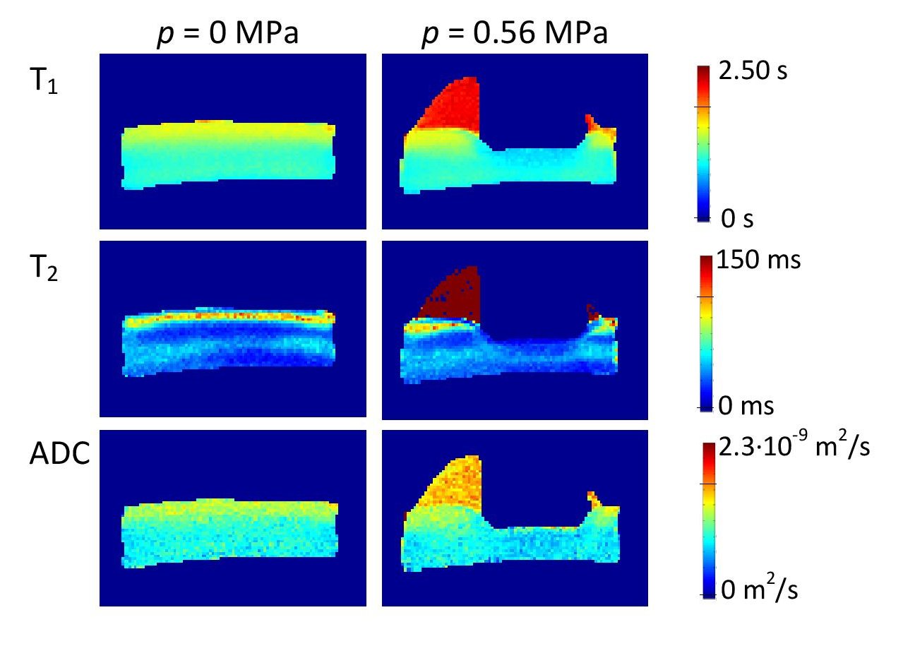
Articular cartilage is a comparatively thin but complex multicomponent avascular tissue, of which main purpose is to provide a low-friction and force-distributing material between the joint surfaces of subchondral bones. Articular cartilage consists of an extracellular matrix, which is composed of a collagen fiber network with intermeshed bottlebrush-like assemblies of proteoglycan molecules, of embedded chondrocytes as well as of water molecules in the interstitial vacancies. Due to depth-dependent orientation of collagen fibers, articular cartilage exhibits three clearly distinguishable layers, i.e., the radial zone close to the cartilage-bone interface, the intermediate zone and the tangential zone at the articular surface. In the study, bovine cartilage-on-bone samples ex vivo were dynamically imaged during their gradual compression up to 0.6 MPa using the FLASH sequence. After complete viscoelastic equilibration, each sample was examined also by multiparametric MR microscopy [1] consisting of inversion recovery SE sequence for T1 mapping, CPMG magnetization-prepared for T2 mapping, diffusion weighted imaging for ADC mapping, diffusion tensor imaging as well as magnetization transfer MR microscopy. Indentation experiments were conducted at 7.0 T using a compression load cell. Spatial resolution of 156 µm x 156 µm x 2000 µm enabled discrimination between compressed cartilage tissue in the compression zone and the intact cartilage tissue. Dynamical imaging revealed gradual compression-induced dehydration of articular cartilage during its compression. Protein densification due to water redistribution from the compression zone to the articular cartilage was consistently confirmed also by multiparametric MR microscopy. The applied experimental approach was found efficient for assessment of the viscoelastic response of articular cartilage as well as of its compression-induced structural changes.

