Examining porous media by correlating internal gradients and pore size distributions
- State Key Laboratory of Petroleum Resource and Prospecting, China University of Petroleum, Beijing, China
An internal magnetic field gradient Gint arises from the magnetic susceptibility difference between different phases in an object. The strength of the internal gradient is approximately proportional to the strength of the applied magnetic field and inversely proportional to the radius of the pore. Due to the fact the internal gradient relates to the pore geometry, distributions of internal gradients can be used to characterize pore structure in porous media [1-3]. So it is desirable to obtain the information about internal gradients. However, internal gradients are really complex in porous media.
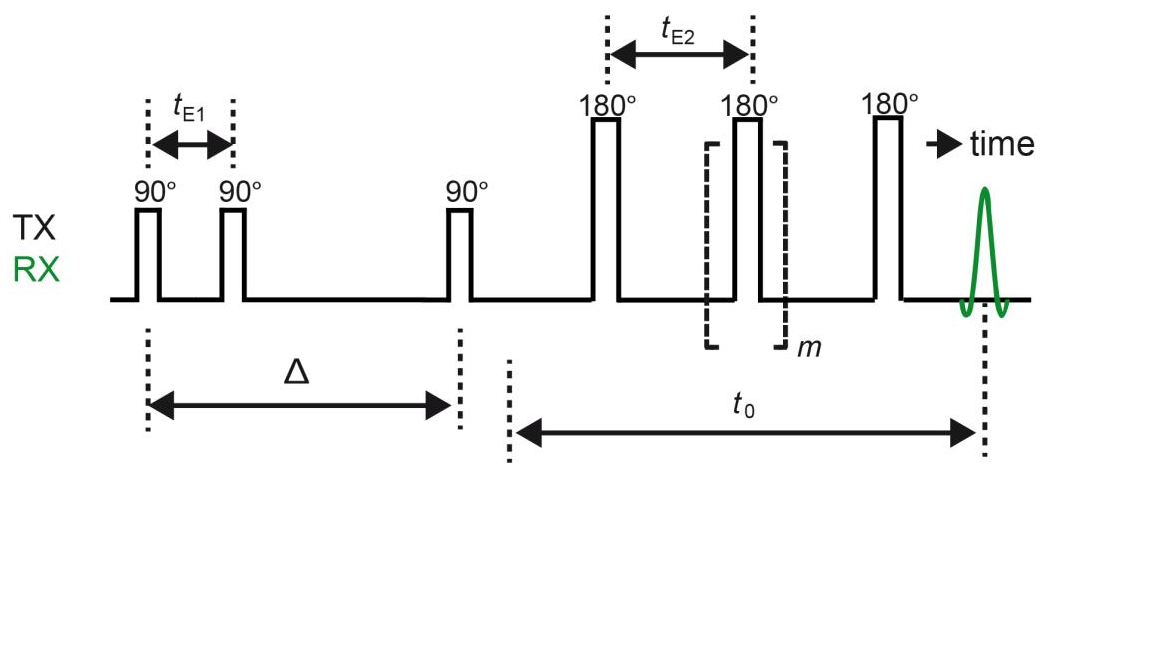
Here we present a new method which correlate internal gradient distributions to pore size distributions. The pulse sequence is divided into two parts as shown in Fig. 1. The first part of the pulse sequence measures the pore size distribution by using Decay due to Diffusion in Internal Field (DDIF) method [4]. The big delta, Δ, was varied Logarithmically from 1 ms to 1 s. The second part of the pulse sequence measures the internal gradient distribution. The information was encoded by varying the number m of 180o pulses from 1 to 100 in 30 steps with constant duration t0 = 60 ms. T1 effect was eliminated by subtracting reference data from the data obtained by pulse sequence shown in Fig. 1. The internal gradient - pore size distribution correlation maps were obtained for glass-bead and rock core samples. Then the relationship between internal gradients and pore structure was analyzed in detail by considering restricted diffusion behavior of fluids in porous samples. Diffusion regimes for each case were assigned by calculating and comparing the diffusion lengths, the dephaseing lengths and the lengths of the pore. For the cases where the free diffusion regime is applied the correlation maps reveal the true relationship between pore structure and internal gradients. And the Δ can be extracted from the correlation maps for the samples having a wide range of pore size distribution. The experiments were implemented on 2 MHz Rock core analyzer (Magritek) and 20 MHz home-made Halbach magnet with Kea2 spectrometer (Magritek). The influence of applied magnetic field on internal gradient and the correlation maps were explored. This method provides useful information about porous media and an insight about the pore structure and internal gradient.

- [1] K. E. Washburn, C. D. Eccles, P. T. Callaghan, (2008), The dependence on magnetic field strength of correlated internal gradient relaxation time distribution in heterogeneous materials, J. Magn. Reson., 33-40, 194
- [2] B. Sun and K. -J. Dunn, (2002), Probing the internal field gradients of porous media, Phys. Rev. E, 051309, 65
- [3] Y.-Q. Song, S. Ryu and P. N. Sen, (2000), Determining multiple length scales in rocks, Nature, 178-181, 406
- [4] H. Liu, M. N. d'Eurydice, S. Obruchkov, P. Galvosas, (2014), Determining pore length scales and pore surface relaxivity of rock cores by internal magnetic fields modulation at 2 MHz NMR, J. Magn. Reson., 110-118, 246
