Multiparametric MRI characterization of magnetic viral complexes
- 1. Technische Universität München, Zentralinstitut für Medizintechnik, Garching, Germany
- 2. Klinikum rechts der Isar der Technischen Universität München, Department of Diagnostic and Interventional Radiology, Munich, Germany
Magnetic nanoparticles (MNPs) can be used as carriers for magnetic drug targeting [1]. When assembled with oncolytic viruses they are a promising tool for cancer treatment. Spatial distribution of particle concentration and aggregation state of the MNP-virus-complexes in the tissue of interest can be assessed by MRI relaxometry allowing for non-invasive therapy monitoring. We investigated the influence of the assembling of MNPs with adenoviruses (Ad) on MRI relaxation rates.
PEI-Mag2 nanoparticles were assembled with oncolytic adenovirus as described in [2]. Dilution series of free particles and magnetic viral complexes were prepared and measured on a 3 Tesla whole body imaging system. R1, R2 and R2* maps were measured using spin echo inversion recovery, spin echo and multi-echo gradient echo sequences. Signal intensities were least-squares fitted for each voxel.
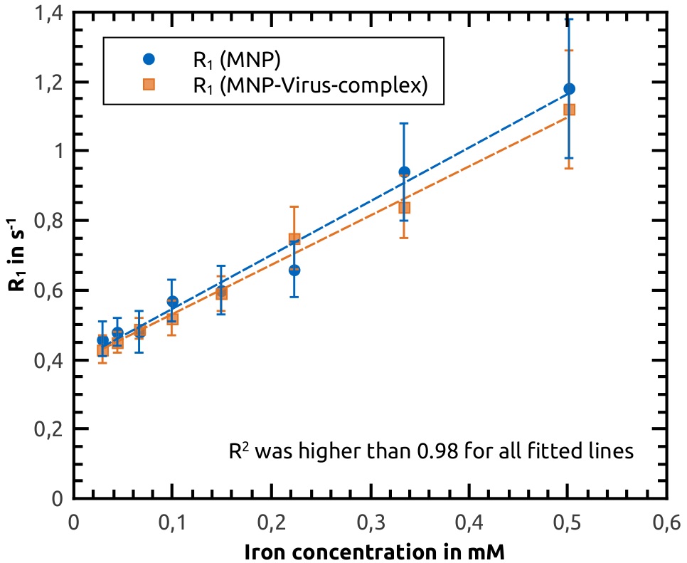
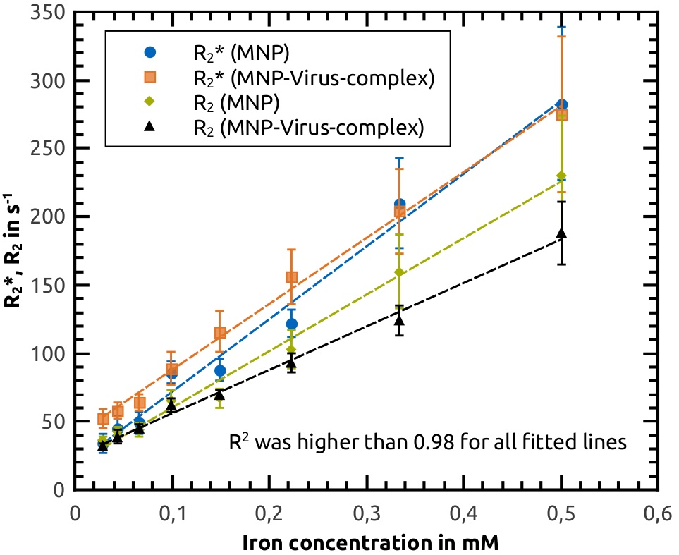
The figures show the relaxation rates of the dilution series with linear fits. Relaxivities for the free particles were r1 = 1.5 ± 0.1 mM-1s-1, r2 = 414 ± 18 mM-1s-1, r2* = 532 ± 26 mM-1s-1 and for the magnetic viral complexes r1 = 1.45 ± 0.05 mM-1s-1, r2 = 318 ± 10 mM-1s-1, r2* = 483 ± 14 mM-1s-1.
The r1 and r2* relaxivities do not change significantly within the error margins when the particles form complexes with the viruses. Therefore R2* relaxation rates seem best suited for particle quantification after a calibration step, whereas R1 rates are too low for good contrast. However, r2 relaxivity decreases when the complexes are formed, probably because of partial refocusing [3]. This indicates, that r2 measurements can be used to determine the aggregation state of the complexes.
- [1] Pankhurst Q A et al., (2009), Progress in applications of magnetic nanoparticles in biomedicine, J Phys D: Appl Phys 42, 224001
- [2] Almstätter I et al., (2015), Characterization of Magnetic Viral Complexes for Targeted Delivery in Oncology, Theranostics 5, 667-685
- [3] Roch A et al., (2005), Superparamagnetic colloid suspensions: Water magnetic relaxation and clustering, J Magn Magn Mater 293, 532-539