Concurrent Optical and MR Microscopy
- 1. University of Freiburg, IMTEK, Laboratory for Microactuators, Freiburg, Germany
- 2. University Medical Center Freiburg, Medical Physics, Freiburg, Germany
We present an MR-compatible optical microscope that can observe samples inside the MR scanner (in contrast [1],[2]) in real-time during MR acquisition with plan-apochromatic aberration correction and a high aperture for selective focusing. The system is inserted with the sample into a small animal scanner where the MRI is obtained using a quadrature coil or an integrated micro coil. This combined imaging may help to identify the object and possible artefacts and to correlate optical and MR contrasts, in particular for (e.g. in-vivo) samples that are not fixated. It can further support dynamic imaging and, potentially, motion correction.
The aims were configurable 0.5, 1 or 2mm fields of view at a resolution of 0.5, 1 and 2µm, respectively; transverse focusing and RGBW top and bottom, dark and bright field illumination. In addition, the used Bruker BioSpec 94/20 gives a maximum 70mm diameter (from the quadrature RF coil) with a transverse observation direction (for RF micro coils) and the gradient, RF and 9.4T B0 field.

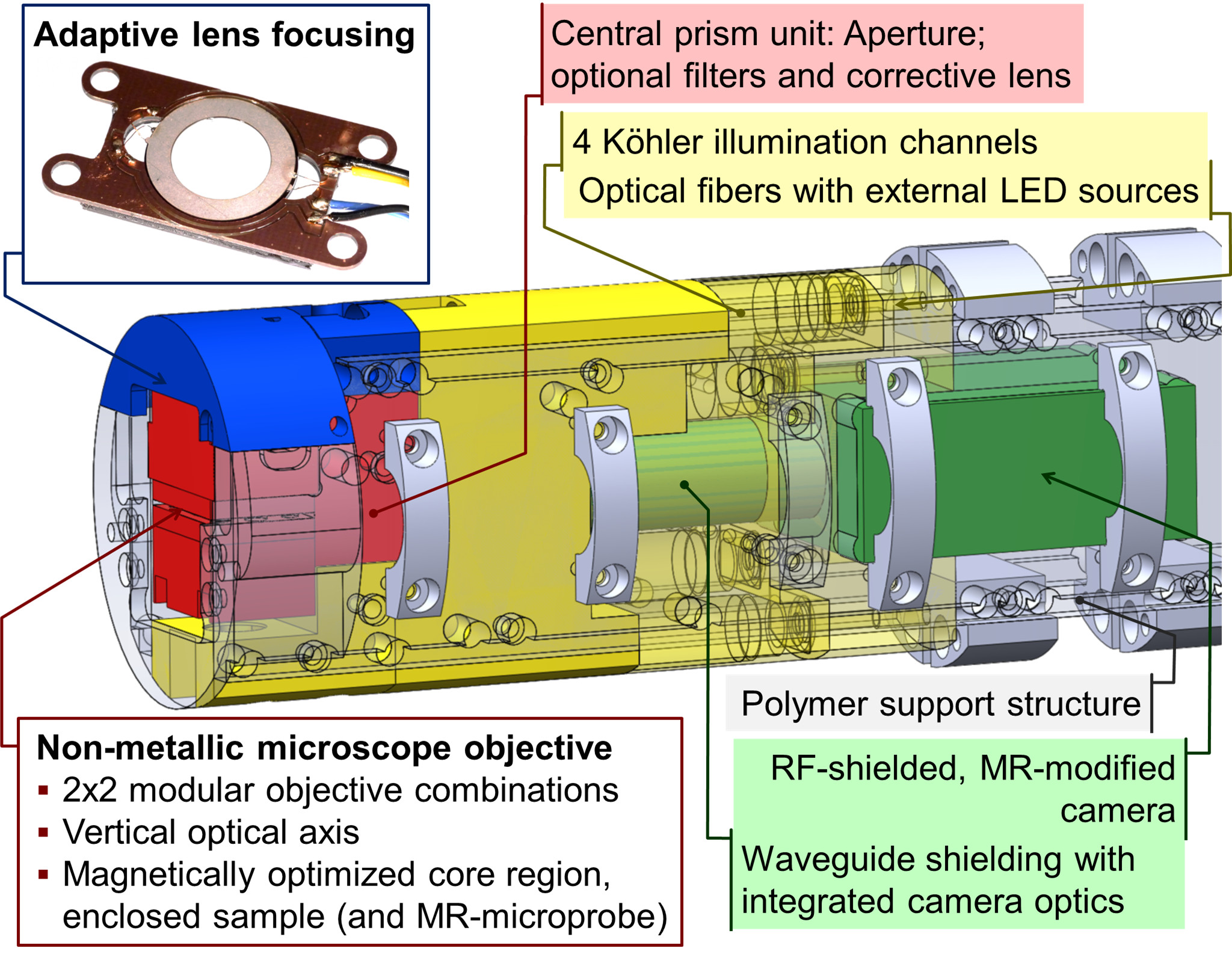
The system uses custom immersion microscope objectives (f=8.4 and f=15.6mm) built from polymer, a multiply tilted (90°) optical path. The magnetic optimization near the sample is based on N-SF11 glass and polyurethane [3], an optimized geometry and immersion in PFPE or halocarbon oil. To minimize motion and metal content, the focusing uses a piezo-driven adaptive lens [4], and the illumination uses fiber-coupled external LEDs. The camera is located on the axis of the magnet, magnetic parts were removed and it uses a waveguide tube for RF-screening with integrated camera objectives (f=25mm and f=50mm).
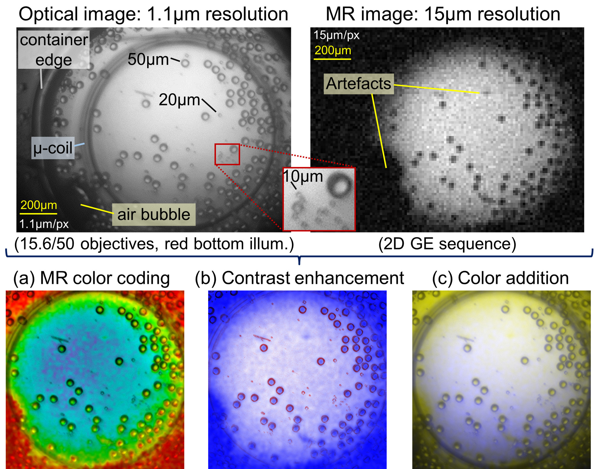
The measured B0 field inhomogeneity using a 3D GE sequence was 19ppb RMS; the RF noise increase was 0.5%. No artefacts were found in the optical image.

For high-resolution MRI, we integrated a Helmholtz micro-coil[5] with single-use PMMA sample carriers and a Bruker micro-coil. For demonstration, we show a mixture of glass beads where we see the higher optical resolution, the identification of artefacts and combined visualizations.
Funding: Baden-Württemberg Stiftung ADOPT-TOMO and BrainLinks-BrainTools DFG EXC 1086.
- [1] Meadowcroft, M. D., Zhang, S., Liu, W., Park, B. S., Connor, J. R., Collins, C. M., Smith, M. B. and Yang, Q, (2007), Direct magnetic resonance imaging of histological tissue samples at 3.0 T, Magnetic Resonance in Medicine 57, no. 5, 835-841
- [2] Demyanenko, A. V., Nie, S, Kee, Y., Bronner-Fraser, M., and Tyszka, M., (2011), Dual-Mode Optical-MR Microscopy with Uniplanar Gradient Coils, ISMRM 2011, Abstract #38
- [3] Wapler, M. C., Leupold, J., Dragonu, I., von Elverfeld, D., Zaitsev, M., and Wallrabe, U., (2014), Magnetic properties of materials for MR engineering, micro-MR and beyond, Journal of Magnetic Resonance 242, 233-242
- [4] Wapler, M. C., Stürmer, M., and Wallrabe, U., (2014), A compact, large-aperture tunable lens with adaptive spherical correction, IEEE International Symposium on Optomechatronic Technologies , arXiv:1411.3746
- [5] Spengler, N., Moazenzadeh, A., Meier, R. C., Badilita, V., Korvink, J. G., & Wallrabe, U., (2014), Micro-fabricated Helmholtz coil featuring disposable microfluidic sample inserts for applications in nuclear magnetic resonance, Micromechanics and Microengineering 24, no. 3 , 034004
