NMR microimaging of growth process of rhizome axillary bud
- 1. University of Tsukuba, Institute of Applied Physics, Tsukuba, Japan
- 2. Tohoku University, Graduate School of Life Sciences, Sendai, Japan
Rice is the principal cereal in the human diet. The red rice, Oryzma longistaminata, is a rhizomatous perennial wild species. The rhizome is an essential, underground stem which enables species to invade new areas, thus allowing overwintering and rapid growth at the beginning of the next growing season. The axillary bud, an embryonic shoot lying at the node in the stem, plays an important role in the rice growth. The axillary bud has developmental mechanisms of dormancy and outgrowth which are regulated in response to environmental cues. However, the axially bud consists of multiple layers of cells and no direct observation of the inner structures has been reported. Here, the anatomical structures of living axillary buds was observed in situ by NMR microimaging, which will provide valuable data to our understanding of rhizome development and function.
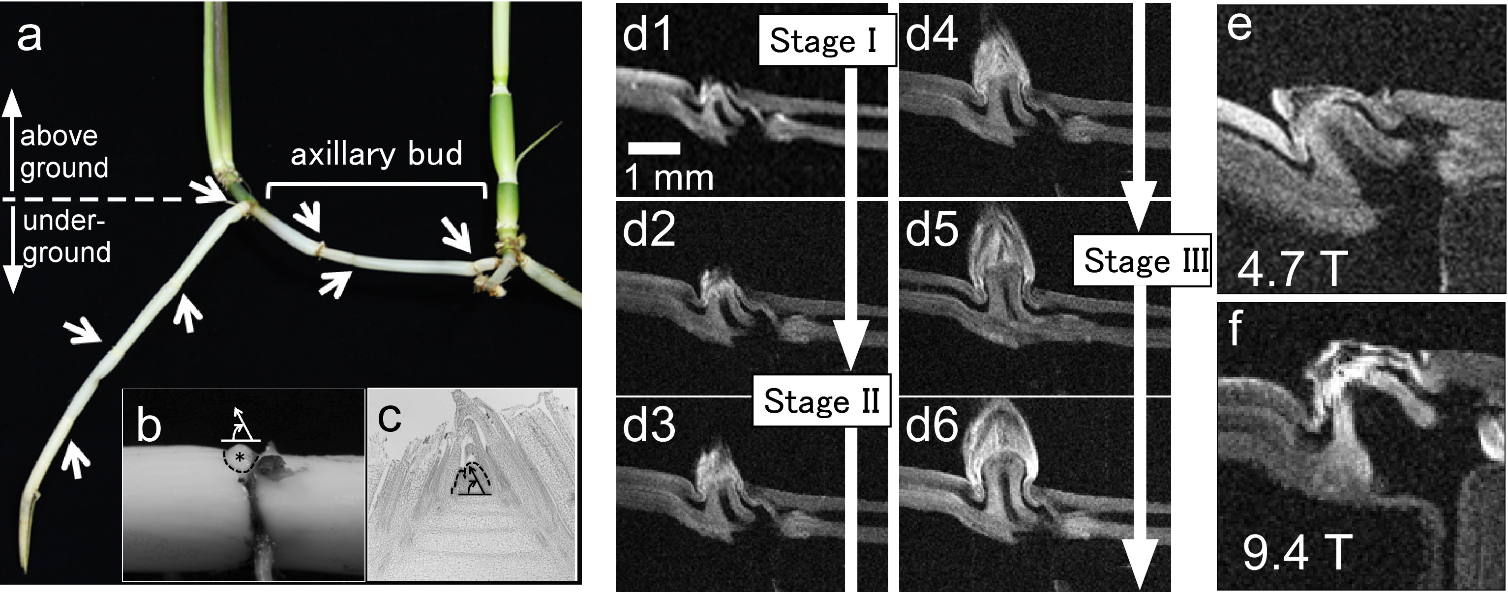
Rhizome axially buds of Oryza longistaminata (Fig. a-c) were observed using 4.7 T/89 mm and 9.4 T/54 mm vertical-bore superconducting magnet systems. For imaging, a part of stem including a bud was cut (3 cm long), terminated with moistened cotton wools, and placed in a sample tube (10 mm in diameter). For both NMR systems, homebuilt solenoid RF probes (12 mm in diameter) were used. For the 4.7 T system, planar gradient coils with the gaps of 26-31mm were constructed, and the efficiency for x, y, and z-gradients was 46.2, 37.8, and 69.0 mT/m/A, respectively. For the 9.4 T system, cylindrical gradients were used and the efficiency was 26.5, 26.9, and 34.6 mT/m/A, respectively. The spin echo sequence (TE/TR = 8 ms/200ms) was used for imaging.
Figures d1-6 show sequential MR images of a growing bud obtained using the 4.7 T system. Although the bud sample was imaged in vitro, it was observed to grow in the same manner as a natural bud does; it underwent a period of dormancy and three developmental stages (I, II, and III). The anatomical structures of the bud including the vascular bundles and the meristem containing undifferentiated cells (stem cells) were sufficiently visualized, especially at the later stages (II and III). The interesting features such as a change in the growth direction of the bundles were observed. However, the spatial resolution was not so high enough to resolve the detailed structures at the initial stage I where the typical dimension of cells is 10-20 μ m. The image signal-to-noise ratio (SNR) was high for the 9.4 T system (SNR = 11.9) compared with the 4.7 T system (SNR = 6.4) (Figs. e and f), showing the potential for resolving the more detailed anatomical structures.

