Examination of menisci in high and low magnetic fields
- 1. RWTH Aachen University, Institut für Technische und Makromolekulare Chemie, Aachen, Germany
- 2. Aachen University Hospital, Department of Orthopaedics, Aachen, Germany
Injuries of menisci are one the most common disease patterns in orthopedics. People suffer from injuries of the meniscus at different ages, due to intensive sport activities, exposure of the knee at work or degenerative processes caused by aging. Every knee contains two menisci, the ring shaped outer meniscus and the half moon shaped inner meniscus. They adjust the incongruity of the femur (thighbone) and tibia (shinbone), leading to a reduction of the joint pressure. [1]
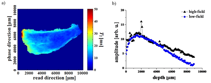
The possible applicability of a low-magnetic field device, the NMR-MOUSE® (Mobile Universal Surface Explorer), in orthopedics was demonstrated. [2] Measurements were conducted at 0,47 T with the NMR-MOUSE® and at 11,7 T/ 16,4 T with a Bruker DSX 500 and a Bruker AVIII 700. The high-field measurements were conducted with the MSME-sequence (Multi Slice Multi Echo) while for low-field measurements the CPMG-sequence (Carr-Purcell-Meiboom-Gill) was used (s. Fig. 1a). For the correlation of the obtained high- and low-field data, T2- and amplitude-profiles were generated and compared. The mean deviation of the correlation of all amplitude-profiles and T2-profiles is approx. 11 %. The deviation may appear large, but the overall trend of the profiles is very similar (s. Fig. 1b).

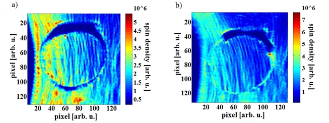
The healing of meniscal tissue injuries is a current topic in medicine with a multitude of different approaches. The possible self-healing potential of meniscal tissue was examined with MRI, as there is no study available which utilizes the benefits of ultra-high magnetic fields. For this study porcine menisci were perforated and analyzed with medically relevant sequences. Using resolutions between 40 and 200 μm/px the changes of the meniscal tissue were examined (s. Fig. 2).

