Dependence of two-phase 19F NMR oxygenation measurements on dispersed-phase droplet size distribution
- 1. Montana State University, Chemical and Biological Engineering, Bozeman, United States
- 2. Montana State University, Center for Biofilm Engineering, Bozeman, United States
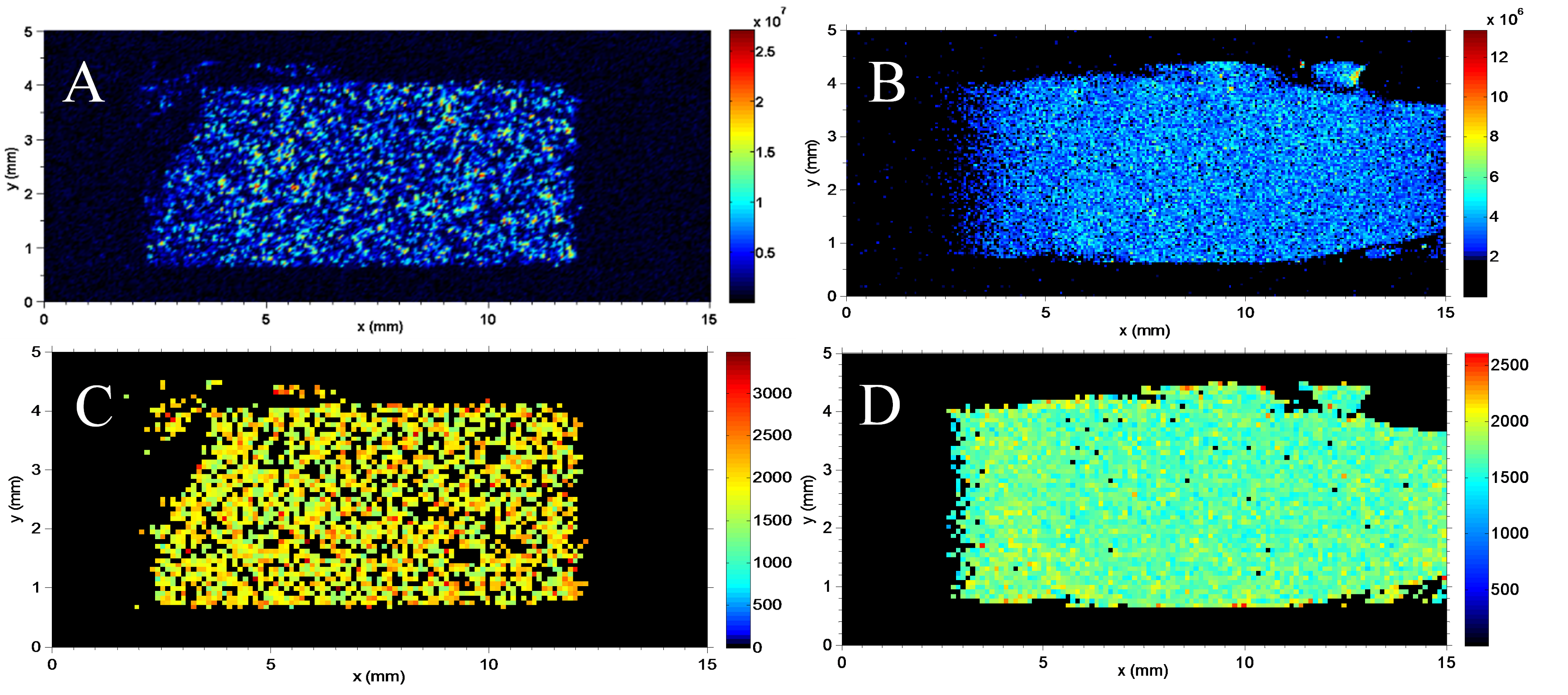
19F magnetic resonance has been explored in the context of medicine as a technique for quantifying oxygenation in blood, tissues, and tumors [1]. The technique is based on the oxygen affinity of fluorocarbons, such as hexafluorobenzene (HFB), that exhibit linear dependence of R1 on local oxygen concentration. As HFB is immiscible in water, it partitions in tissues when injected and the impact of susceptibility artifacts at interfaces on T1 measurements from dispersed HFB in a continuous aqueous phase may exhibit dependences on droplet size [2], and thus not directly correlate with measurements from a single HFB phase, leading to error in oxygenation determination. To evaluate this effect, the impact of HFB droplet size on spatially-resolved T1 measurements was analyzed using agarose gel as an aqueous phase surrogate for tissue. Gel samples of various HFB concentrations were made and the two phases were homogenized with a shear mixer at 5,000 rpm (low-dispersion) or 15,000 rpm (high-dispersion) to generate different droplet size distributions. T1 inversion recovery imaging revealed a strong dependence of spatially-resolved T1 distribution on mean droplet radius, with smaller droplets displaying higher T1 values. Our results suggest that applying a standard curve generated from single-phase T1 measurements to measurements from a dispersed HFB phase may result in systematic overestimation of local oxygenation. This research is a first step toward developing a technique to measure the spatiotemporal distribution of oxygen in clinically relevant biofilms such as those of Staphylococcus aureus.

- [1] Mason, R. P., Nunnally, R. L., Antich, P. P., (1991), Tissue oxygenation - a novel determination using 19F surface coil nmr spectroscopy of sequestered perfluorocarbon emulsion, Magnetic Resonance in Medicine 18, 71-79
- [2] Blinc, R., Dolinsek, J., Lahajnar, G., Sepe, A., Zupancic, I., Zumer, S., Milia, F., Pintar, M. M., (1988), Spin-lattice relaxation of water in cement gels, Zeitschrift für Naturforschung A 43(12), 1026-1038
