Visualization of water flow in mantle cavity of bivalve by 7T MRI
- 1. Dokkyo Medical University School of Medicine, Regulatory Physiology, Shimotsuga-gun, Japan
- 2. Atmosphere and Ocean Research Institute, University of Tokyo, Division of Marine Life Science, Kashiwa, Japan
- 3. National Institute for Physiological Sciences, Okazaki, Japan
- 4. Japan Agency for Marine-Earth Science and Technology, Institute of Biogeosciences, Yokosuka, Japan
Water flow inside shells of bivalves (Mytilus galloprovincialis) was measured by phase-contrast magnetic resonance imaging (PC-MRI), and transient changes in water velocity were imaged by using the inflow effect of T1-weighted MRI (Fig. 1A,B). All experiments were done by 7 T high-field MRI (Avance III 300wb, Bruker) at 293 K. The mantle cavity inside the shells is separated into upper and lower mantle cavities by a pair of W-shaped gill. During steady ventilation (Fig. 1C), water entered from the inhalant aperture (40-20 mm s-1), and filled in the lower mantle cavity. Then, water approached to the demibranches of gill (5-10 mm s-1). After passing through the lamellae to the upper mantle cavity, the water stretched the interlamellar cavity, and turned to posterior direction in the upper mantle cavity, and exhaled out from the exhalant siphon (50 mm s-1). Spontaneous opening of the shells caused a quick increase of the flow in the mantle cavity within 1 min (Fig. 2). There was a high correlation between the area in the inhalant aperture and that in the exhalant siphon. However, the flow in the interlamellar cavity showed a low correlation with that in the inhalant aperture. The flow in the right and left interlamellar cavities changed independently. These results suggested that the mussel controls the amount of flow by changing the area and region of active lateral cilia in the demibranches. In conclusion, a combination of PC-MRI and the inflow effect of T1w-MRI allowed us to perform quantitative flow analysis in all of the cavities in the mussel.
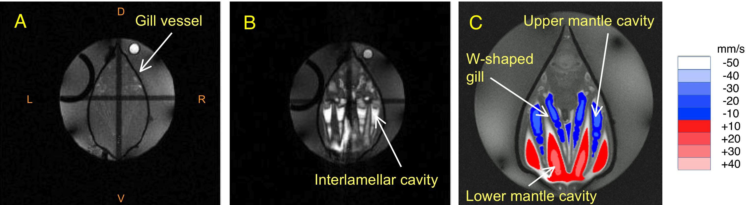
Fig. 1 Transverse T1w-MRI of M. galloprovincialis at resting condition (A), and during ventilation (B). Anterior-posterior flow measured by PC-MRI (C). Anterior- and posterior-directed flows of water are shown in red and blue, respectively.
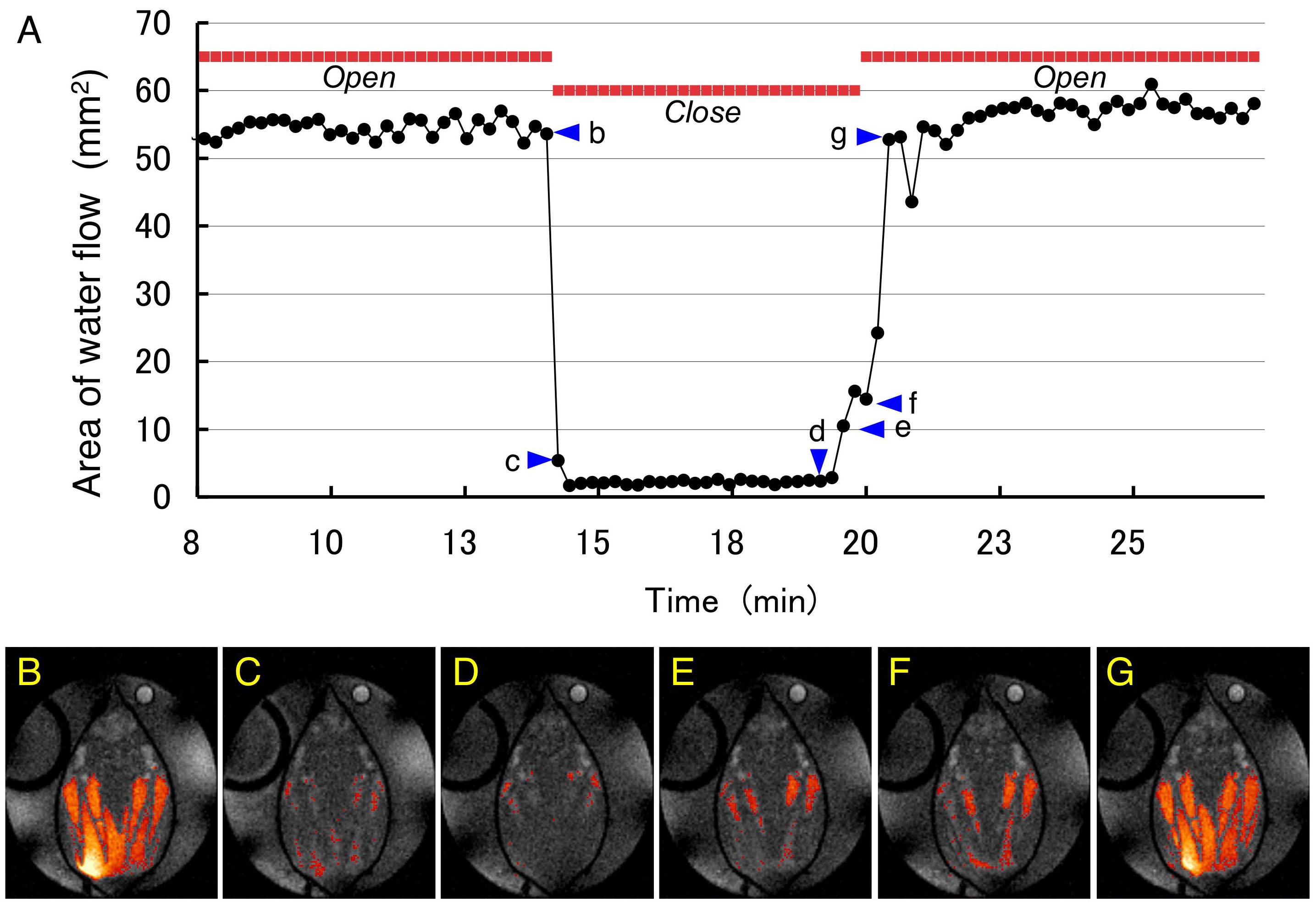
Fig. 2 Changes in water flow in the mantle cavity of M. galloprovincialis. (A) Changes in the transverse cross-sectional area of the water flow. 'Open' and 'Close' represent the closure and opening of the shell of the mussel, respectively. (B-G) Transient changes in the water flow before/after and closure/open of the shells. The timing of each image is shown by the arrowheads labelled b to g in A. The orange pixels represent water flow.[1]
[1]