Comparative study of Á-MRI and X-ray Á-CT on an oilseed grain
- 1. Karlsruhe Institute of Technology, Institute of Microstructure Technology (IMT), Eggenstein-Leopoldshafen, Germany
- 2. BASF - The Chemical Company, Ludwigshafen, Germany
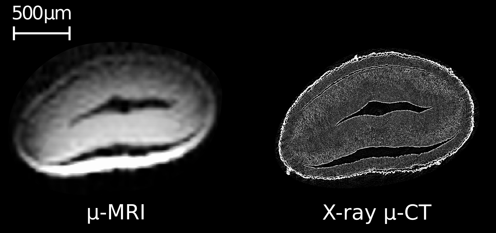
Recent advances in the manufacturing of miniaturized MRI equipment [1] enable MRI with very short dead times and resolutions in the range of 10 Ám. Such imaging conditions are especially interesting for imaging small "hard" biological specimens such as seeds.
With x-ray micro-tomography (ÁCT), resolutions in the range of about 1 Ám or even slightly below can be realized on standard laboratory setups. At the same time, however, the contrast information in the X-ray images is quite rudimentary when it comes to the organic constituents of the seeds.
Due to the availability of both relaxation time and chemical shift as specific contrast moieties to discern oil and water, ÁMRI is much better fitted to provide this specific information on the local composition of the seed. With the considerably shrunk gap in resolution between the two techniques, correlating features found by the two imaging techniques on the same grain comes into the reach of feasibility.
In our contribution, we present imaging results on sesame and rape seed grains along with NMR spectroscopic data obtained on the same seeds.

