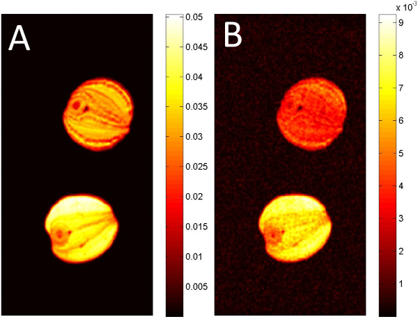
3D distribution of (un)saturated fatty acid components in seeds
- 1. Leibniz Institute of Plant Genetics and Crop Plant Research, Heterosis, Gatersleben, Germany
- 2. University of Würzburg, Experimental Physics 5, Würzburg, Germany
- 3. MRB Research Center for Magnetic-Resonance-Bavaria, Würzburg, Germany
- 4. Pennsylvania State University, Department of Bioengineering, University Park, United States
Introduction: A great variety of oil-bearing crops is available for production of oil and further nutritional products. One element of a healthy diet is sufficient supply with unsaturated fatty acids.
Inspired by [1], in this work we demonstrate a setup for 3D analysis of unsaturated fatty acid component distribution in various seeds. Comparisons between different types of seeds are rendered possible and information about localized fatty acid composition is available
Subjects and Methods: Dry oil-bearing seeds and parts of larger seeds were placed within a glass tube in a custom-built 4 mm inner diameter solenoid coil. A Varian 14.1 T (600MHz) vertical bore NMR scanner was used for imaging. The Spin-Echo sequence includes a water suppression module which allows to suppress the particular fatty acid component. Therefore the sequence allowed selective imaging of specific components of the lipid molecules, i.e. protons located at the saturated or unsaturated carbon atoms respectively. Parameters used: TR 675 ms, TE 4.5 ms, FoV 8 x 4 x 4 mm³, Matrix 192 x 96 x 96, Resolution 41.7 µm isotropic. Number of averages: saturated components NA=4, unsaturated components NA=8. Overall scan time 20h 44m.

Results and Discussion: The 3D datasets show the individual component distribution of the particular seeds. In Fig.1 two different types of rapeseed canola seeds are compared, clearly showing their different overall lipid content (Fig. 1A, B). The calculated ratio-map (data not shown) reveals the higher ratio of unsaturated fatty acid components in the bottom seed.
In the presentation we will show the individual ratios of "saturated : unsaturated "fatty acid components of pine nut, coconut-meat and olive fruit. Even though Coconut and Pine-Nut seem to have identical signal-intensities concerning their (unsaturated) lipid signal in direct comparison, they differ considerably in their lipid component ratios and therefore in their healthful characteristic.
Conclusion: In this work we present a method for comparison of different oil-bearing seeds regarding their content of unsaturated lipid components. Significant differences in ratios of unsaturated versus saturated components of lipids can even occur within the same type of seed and can easily be detected with this technique.
