Seeing the unseen in plants with UTE-imaging
- 1. Leibniz Institute of Plant Genetics and Crop Plant Research, Heterosis, Gatersleben, Germany
- 2. University of Würzburg, Experimental Physics 5, Würzburg, Germany
- 3. MRB Research Center for Magnetic-Resonance-Bavaria, Würzburg, Germany
Introduction:
Imaging plants can be challenging, i.e. due to susceptibility changes within the plant tissue or when using dry seeds as object of interest, which have short T2 relaxation times impeding MR-imaging [1] , [2] . In this work we demonstrate that ultra-short echo-time (UTE) imaging affords new opportunities to examine plant structures of materials typically invisible in conventional MRI-sequences (e.g. dry seeds).
Subjects and Methods:
We used a custom UTE-Sequence on a Bruker 11.7 T (500 MHz) vertical bore NMR scanner. Dry seeds were placed in a 5 mm inner diameter glass tube and placed in a home-built 5 mm Helmholtz-coil. Spin-Echo images were acquired for comparison with a short echo Spin-Echo (SE) Sequence. SE-Parameters: TR 500 ms, TE 4.43 ms, FoV 10 x 5 x 5 mm³, Matrix 96 x 48 x 48, 6 averages, 2 repetitions. Resolution 104µm isotropic. UTE-Parameters: TR 100 ms, Acquisition-Delay 10 µs, cone-based trajectory for anisotropic FoV (15 x 5 x 5 mm³) [3] , projections 58772, Readout-points 128, cylindrical trajectory, 6 averages, 2 repetitions. Resolution 100 µm isotropic.

Results and Discussion:
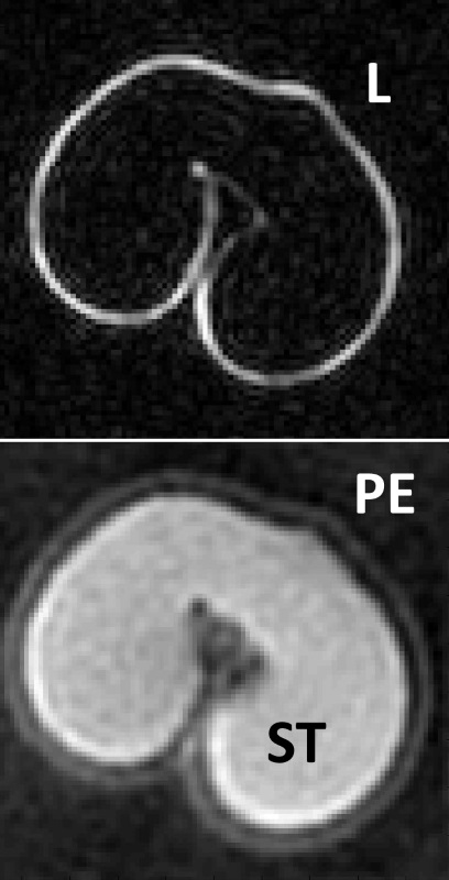
Imaging of dry wheat grain with a SE-sequence is usually restricted to the lipid signal originating mostly from the embryo und a small layer close to the pericarp. By using the ultra-short echo-time sequences, even the signal from the endosperm (mostly starch protons) can be acquired. In Fig. 1 also the seed's coat is detectable in the images. Variations of the Echo-Delays showed a considerable loss of signal beyond 250µs.
Further experiments (plant stems, siliques, spikes) confirmed the advantages in plant imaging gained by the UTE sequence.
Conclusion:
Standard Sequences (e.g. Spin-Echo) suffer great signal loss within tissues with short T2-times. Since this leads to a deteriorated image quality, a large amount of information is therefore inaccessible. As shown above UTE-imaging enables imaging of dry materials (seeds) as well as tissues with air inclusions (living plants). The internal structure of seeds can be visualized and volumes of the seed's organs can be calculated.
- [1] Borisjuk and Melkus, et al., (2014), In vivo imaging: new researc, Nova Science Publishers, New York, 1-48, ISBN 978-1-62948-633-8
- [2] Stapley et al, (1997), NMR imaging of the wheat grain cooking process, Blackwell Science Ltd, International Journal of Food Science and Technology, 355-375
- [3] Larson et al, (2008), Anisotropic Field-of-Views in Radial Imaging, IEE Trans. Med. Imaging, 47-57
